| Tour 1: Next/Previous/Start:
The following section summarizes the brain damage in
this case,
and links each structure mentioned to its entry in the
Atlas of the normal brain.
Move through the dataset (use the buttons at left) to view each of the structures mentioned.
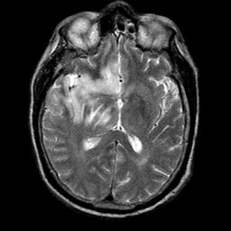
Abnormally high signal extends from the right temporal tip medially to
involve the hippocampus,
amygdala, and
the parahippocampal gyrus,
superiorly to involve
orbital frontal gyri, the
straight gyrus, and the
cingulate gyrus; and posteriorly and superiorly to involve the insula, the anterior
bank of the superior
temporal gyrus, and the posterior
hippocampus as it blends into the column of
the fornix.
Medially, the lesion displays evidence of tracking along an anatomically
restricted course, as it borders the posterior genu of
the
internal capsule. The greatest superior
extent of the lesion involves the posterior portion of the medial
nucleus of the thalamus,
a structure
known to have anatomic connections with
amygdala and other limbic structures (Nauta WJH. Neural association of
the amygdaloid complex in the monkey. Brain 1962; 85:505-520).
HSV has also damaged the intralaminar thalamic nuclei,
structures known to
have extensive connections with the limbic system (Van
Landingham KE and Lothman EW. Self-sustaining limbic
status epilepticus. Neurology 1991;41:1942-9.)
|