| Tour 1: Next/Previous/Start:
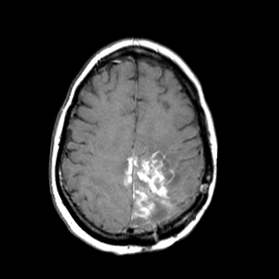
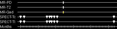
The core of this lesion contains elements which enhance with gadolinium,
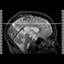
as seen here at the same slice location as before. Choose a spatial movie
of this data (by choosing "cine" next to the sagittal image), to see the entire extent of gadolinium enhancement. Note that
there is extension of the enhancing portion of the mass to the right hemisphere.
In this T1-weighted series, highest signal comes from water which has had its
relaxation time shortened because of proximity to molecules of the injected
contrast agent gadolinium-DTPA. Such signal is generally seen from water
which is either intravascular or within regions of breakdown of the blood-brain barrier.
|
|