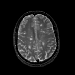
| Tour 1: Next/Previous/Start: Now look at the temporal movie at this level, and you will see three
lesions of approximately the same size, which appear and
nearly disappear, along with the associated halo of
edema. The 3 foci of inflammitory activity are clearly not in synchrony.
Notice the left lower lesion: it is at the base of the marginal
sulcus and occupies white matter under the right post-central
gyrus. As the acute inflammation grows, notice how the architecture
of the post-central sulcus is displaced posterolaterally. Near the
end of the year, this lesion has almost disappeared, but another
has appeared just behind it, in the slice below. In
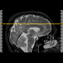
Tour #2, we will consider a lower, periventricular slice,
in which a large range of lesion size is seen.
|
|