 |
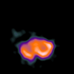
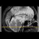
| Tour 2: Next/Previous/Start: As you can see from the yellow line cursor in the sagittal locator below, this slice is located in the mid-portion of the cerebellum. The Tc99-HMPAO uptake at this level is much higher that of either the frontal or the parietal lobes. Note that much of the cerebellar grey matter is white, indicating high perfusion. |
|
|
|||||||||||
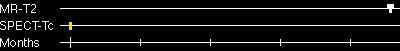
| [Home][Help][Clinical][Tour 1][Tour 2] | Slice 9 |
| Click on sagittal image to select slice. Click on thin tickmark to change timepoint, or thick tickmark for overlay. | |
| Keith A. Johnson (keith@bwh.harvard.edu), J. Alex Becker (jabecker@mit.edu) | |