 |
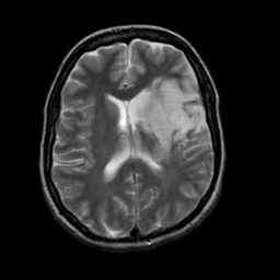
| Tour 1: Next/Previous/Start: Note how the conventional T2-weighted images have converted to high signal in the lesion at four days after onset of symptoms. Compare these images with the diffusion-weighted MR images obtained acutely (below) |
|
|
|||||||||||||
| [Home][Help][Clinical][Tour 1] | Slice 15 |
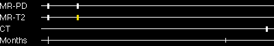
| Click on sagittal image to select slice. Click on thin tickmark to change timepoint, or thick tickmark for overlay. | |
| Keith A. Johnson (keith@bwh.harvard.edu), J. Alex Becker (jabecker@mit.edu) | |