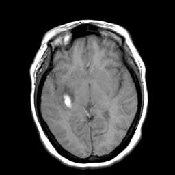
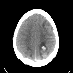
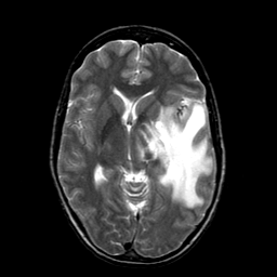
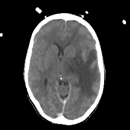
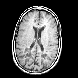
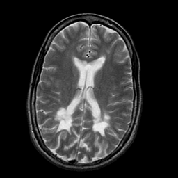
Normal tissue
| MR-T11 | MR-T21 | xray-CT2 | |
| dense bone | dark | dark | bright |
| air | dark | dark | dark |
| fat | bright | bright | dark |
| water | dark | bright | dark |
| brain | "anatomic"3 | interm. | interm. |
1. Bright means high signal intensity, dark means low, and interm. means intermediate.
2. Bright means high density/high attenuation of x-rays, dark means low.
3. Grey matter appears grey, white matter white.
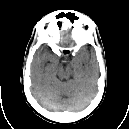
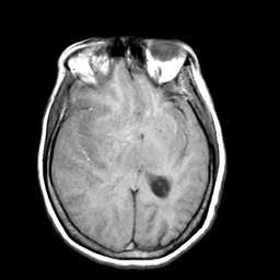
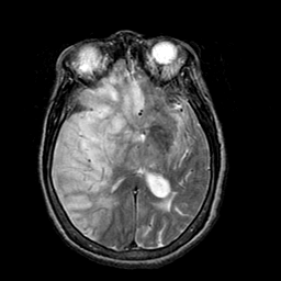
Abnormal tissue
| MR-T1 | MR-T2 | xray-CT | enhancement1 | |
| infarct | dark | bright | dark | subacute |
| bleed | bright2 | bright2 | bright | no |
| tumor | dark | bright | dark3 | yes |
| MS plaque | dark | bright | dark4 | acute |
1. Blood brain barrier leak. For MR, gadolinium; for CT, iodinated contrast material.
2. Unless very fresh or very old.
3. Unless calcified.
4. Often isodense.
Interpretation of neuroimaging should be performed by qualified professionals.