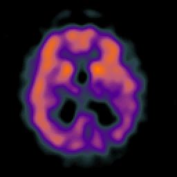
| Tour 2: Next/Previous/Start: Here is a mid-ventricular slice which demonstrates the commonest
finding in functional imaging of Alzheimer's disease. (Check the
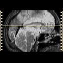
corresponding anatomic image by choosing the MR-T2 tickmark
on the timeline, or by using the arrow buttons at right)
The dark blue regions in the parietal lobes represent areas of
decreased blood flow or perfusion. This reduction in blood flow is
due in part to the underlying atrophy, in part to the presence of diseased
brain, and in part to the functional
"disconnection" of this from other brain regions affected by the disease.
|
|


