|
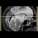
| Tour 1: Next/Previous/Start: At this level, the orbital frontal and superior temporal structures are seen. Note the slight asymmetry of lateral ventricles, with the left posterior portion, sometimes called the atrium, larger than the right. This is a normal variant, and corresponds to the slight asymmetry of the Sylvian fissures. This slight asymmetry can be confirmed by viewing adjacent slices, or by running the spatial movie (choose "cine" next to the sagittal image). |
|
|
|||||||||||
| [Home][Help][Clinical][Tour 1][Tour 2] | Slice 26 |
| Click on sagittal image to select slice. Click on thin tickmark to change timepoint, or thick tickmark for overlay. | |
| Keith A. Johnson (keith@bwh.harvard.edu), J. Alex Becker (jabecker@mit.edu) | |