| Tour 1: Next/Previous/Start:
Herpes Simplex Virus (HSV) encephalitis has its own neuroanatomy.
It tends to attack a part of the brain known as the "limbic system",
a set of interconnected brain structures responsible for
the integration of emotion, memory, and complex behavior.
This disease is important to recognize because there is an effective
drug treatment, acyclovir.
We will see the limbic system on this tour, as shown by the
lesions of a typical case of HSV encephalitis.
HSV is ubiquitous, but fortunately, only 1 or 2 cases per
million infected individuals develop the encephalitis
of HSV each year in the US. It is the most frequently fatal of
all encephalitides.
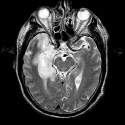
In this set of images, there is a region of very bright signal
on MR (and high blood flow on SPECT; use the buttons at right)
in the medial temporal lobe at left (patient's right). This
corresponds to an area of active viral leptomeningeal and brain
tissue infection. Hemorrhage can occur
acutely, but is not seen in this case. You can see obliteration of
the temporal horn of the
lateral ventricle because of swelling of the hippocampus.
The remainder of the brain is relatively
hypoperfused (use the buttons at right) and structurally
normal. The MR images were obtained 5 days after onset
of symptoms, and the follow-up SPECT 23 days later.
How did this patient's symptoms relate to the location of the lesions? Go to the next tour point.
|


