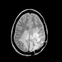
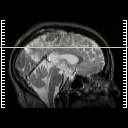
| Tour 2: Next/Previous/Start: This tour will examine the cerebral edema which corresponds generally to the
high signal extending from the center of the mass through surrounding white
matter. On this proton-density-weighted image, the high signal corresponding
to edema respects the gray-white junction, but tends to spare the "u"-fibers.
This is a common observation in the white matter reaction to malignant neoplasia,
so-called "vasogenic" edema. Choose a spatial movie (by clicking "cine" next to
the sagittal image) of the proton density stack
to see the extent of the edema.
|
|