 |
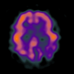
| Tour 2: Next/Previous/Start: Compare the cerebellar slice you just saw to the frontal and parietal lobes in this slice. Both are abnormally reduced in perfusion, consistent with reduced function, but the parietal is clearly the most affected. Note that the left hemisphere is more abnormal than the right. This is a common observation in functional imaging of Alzheimer's disease. |
|
|
|||||||||||
| [Home][Help][Clinical][Tour 1][Tour 2] | Slice 32 |
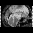
| Click on sagittal image to select slice. Click on thin tickmark to change timepoint, or thick tickmark for overlay. | |
| Keith A. Johnson (keith@bwh.harvard.edu), J. Alex Becker (jabecker@mit.edu) | |


