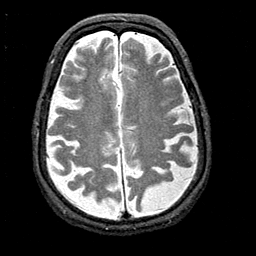
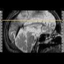
| Tour 2: Next/Previous/Start: Find the central sulcus.
To get properly oriented, view the MPEG
movie ("cine" button, next to the sagittal image) of the entire dataset, and find the central sulcus by first
locating the marginal sulcus in the medial parietal lobe. From
the marginal sulcus, the central is usually the first encountered
when moving anteriorly. Compare
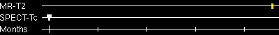
this with the functional image at the same level (use the buttons at right, or
choose the SPECT-Tc tickmark on the timeline). Note that both
pre- and post-
central gyri, where the primary sensori-motor cortices are located, are
relatively hyperperfused. In general, Alzheimer's disease is
associated with reduced brain function, especially in non-primary
regions. The association cortex of the parietal lobes is often
severely affected, as illustrated in this case.
|
|